Instructions for Use of T-Handle Safety Trephine for Spinal Endoscopic Surgery Instruments
1. Product Overview
This instrument is a dedicated T-handle safety trephine for spinal endoscopic surgery, a core supporting tool for minimally invasive spinal surgeries such as percutaneous endoscopic lumbar discectomy (PELD) and spinal endoscopy. It is mainly used to safely and precisely grind/resect hyperplastic bone, calcified tissue, and hypertrophic ligamentum flavum, expand the intervertebral foramen/spinal canal, establish an operative channel for surgery, and minimize the risk of nerve injury.
2. Core Structure and Features
- T-shaped anti-slip handle: Ergonomically designed to provide a stable grip, precisely control the advancement depth and rotation force of the trephine
- Graduated guide rod/trephine shaft: Clear depth markings to real-time monitor surgical operation depth and prevent over-cutting
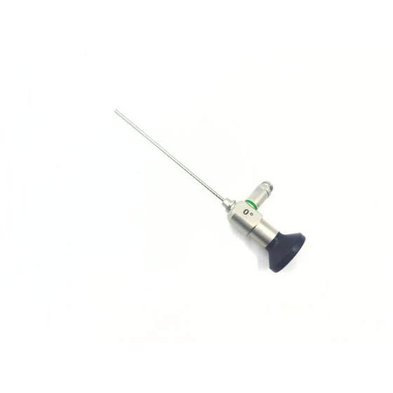
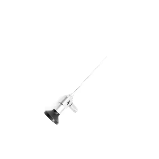
- Safety limit design: Equipped with a safety trephine head to achieve controllable cutting and prevent accidental penetration that may damage the spinal cord/nerve roots
- Medical-grade stainless steel material: Autoclavable at high temperature and pressure, meets surgical sterility requirements, with corrosion resistance and high strength
3. Applicable Surgical Scenarios
- Percutaneous Endoscopic Lumbar Discectomy (PELD): Used for foraminoplasty, grinding hyperplastic facet joints, expanding the intervertebral foramen, and establishing a working channel
- Cervical/Thoracic Spinal Endoscopic Surgery: Spinal canal decompression, intervertebral foramen expansion and plasty
- Minimally Invasive Spinal Decompression Surgery: Resecting calcified intervertebral disc tissue, hypertrophic ligamentum flavum, and hyperplastic osteophytes
- Revision Surgery after PELD: Treating scar tissue and performing secondary foraminoplasty
4. Preoperative Preparation
- Instrument Inspection: Preoperatively check whether the trephine blade is sharp and free of curling, the T-handle connection is firm, and the markings are clear and free of wear
- Sterilization: Use autoclave sterilization (134℃, 3-4 minutes) or ethylene oxide sterilization to ensure a sterile state
- Preoperative Localization: Accurately localize the surgical segment with a C-arm X-ray machine, mark the puncture point and needle insertion path
- Patient Preparation: Routine anesthesia (mainly local anesthesia), position placement (prone position, abdomen suspended)
5. Standard Operating Procedures
- Puncture Localization and Channel Establishment: According to the preoperative plan, use a puncture needle for percutaneous puncture to the target intervertebral foramen, and confirm the position with C-arm fluoroscopy; Insert a guide wire, sequentially place dilators along the guide wire, gradually expand soft tissue, and establish a surgical working channel; Finally place the working cannula, reconfirm the cannula position with C-arm fluoroscopy to ensure a safe operation path for the trephine
- Instrument Installation and Localization: Firmly connect the safety trephine head to the T-handle and check the connection stability; Slowly advance the trephine along the working cannula, aligning with the hyperplastic bone/facet joint to be cut; Confirm the trephine head position with C-arm fluoroscopy, record the initial depth against the markings, and set the safe cutting depth
- Safe Trephine Operation: The surgeon holds the T-handle and rotates it in a slow, uniform, and controllable manner to drive the trephine to cut bone; During operation, real-time check the markings, strictly control the cutting depth, and strictly prohibit exceeding the preoperatively set safety threshold; Intermittently stop operation, confirm the cutting progress with C-arm fluoroscopy to avoid damaging nerve roots and dural sac; Ask the patient's feeling (under local anesthesia), and immediately stop operation and adjust the position if there is radiating pain or numbness in the lower limbs; Adopt the "small amount multiple times" cutting principle to gradually expand the intervertebral foramen/spinal canal until the surgical field of view requirements are met
- Postoperative Instrument Removal and Channel Treatment: After cutting, slowly rotate and withdraw the trephine to avoid the blade catching soft tissue; After removing the trephine, clean bone chips and tissue fragments generated by cutting through the working cannula; Insert a spinal endoscope to check the decompression effect and confirm sufficient nerve release and no residual compression
- Postoperative Instrument Handling: Immediately rinse the instrument with clean water after surgery to remove blood stains and bone chips, focusing on cleaning the trephine blade and handle gaps; Use ultrasonic cleaning, then perform disinfection and sterilization, and store in a sterile instrument cabinet by category
6. Safety Precautions
- Strict Depth Control: Trephine operation must be performed under full C-arm fluoroscopy monitoring, blind operation is strictly prohibited, and markings are the core basis for safe operation
- Nerve Protection Priority: If the patient develops nerve irritation symptoms during operation, stop immediately, adjust the trephine angle/depth, and replace the operation path if necessary
- Instrument Maintenance: Check the blade after each use, replace the trephine head in time if there is curling or wear, to avoid affecting the cutting effect and surgical safety
- Aseptic Operation: Strictly follow the aseptic surgical principle throughout the process to prevent postoperative infection
- Applicable Personnel: Only operated by orthopedic/neurosurgeons qualified in minimally invasive spinal surgery, non-professional personnel are strictly prohibited from using
7. Maintenance and Care
- Cleaning: Clean in time after surgery to avoid blood and tissue residue corroding the instrument
- Sterilization: Strictly follow the medical device sterilization specifications to ensure qualified sterilization
- Storage: Store in a dry and sterile environment to avoid blade collision damage
- Regular Inspection: Regularly check the handle connection, blade status, and marking clarity, and repair/replace damaged parts in a timely manner


-560x560.png)